South Australian Medical Heritage Society Inc
Website for the Virtual Museum
Home
Coming meetings
Past meetings
About the Society
Main Galleries
Medicine
Surgery
Anaesthesia
X-rays
Hospitals,other organisations
Individuals of note
Small Galleries
Ethnic medicine
- Aboriginal
- Chinese
- Mediterran
Electroencephalography (EEG)
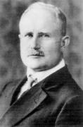
Hans BergerAcknowledgments: We are grateful to Dr. Anne Hamilton-Bruce, Dr. Martin Robinson, Joanna Proszkowsk, members of the Queen Elizabeth Department of Neurology and Ms. Jan Hooper AO of the Division of Medicine. They allowed us to photograph an old and a current EEG machine and provided the relevant information.
The electroencephalogram, (acronym EEG) is a diagnostic tool which records the electrical activity of the brain using numerous electrodes placed on the scalp. The electrical activity is produced by the brain cells (neurones) and neural circuits. It was an important diagnostic aid in the second half of the last century.
The first account of electrical activity in the brain was published by Richard Canton, a Liverpool surgeon in the British Medical Journal in 1875. He recorded electrical activity by placing electrodes on the exposed brains of rabbits and monkeys. Fifteen years later Beck using similar animals noted the changes produced by light and other stimuli.
In 1925 Hans Berger, a German psychiatrist from Jena , recorded brain activity similar to the current recordings using electrodes placed on the human scalp. He named one set of recordings the “alpha” waves and published his findings in 1929. Like Einthoven he used light to record on photographic plates. In 1932 Adrian and Mathews in Cambridge , using copper gauze and saline electrodes, confirmed Berger’s findings, and published their findings in the journal Brain. Berger refused their suggestion to call a set of waves “Berger” waves.
With advances in technology, pen recordings and electrodes were improved and in the 1960s the EEG was an important diagnostic tool. It was used to diagnose strokes, tumours, encephalopathies, brain inflammation, and above all epilepsy. However with the development of modern medical imaging such as CT and MRI , tumours and strokes and other space occupying lesions can be much better demonstrated and located. The main role of EEG now is in the diagnosis of epilepsy, coma, brain death and in excluding an epileptic component in encephalopathies.
The historic information is from Wikipedia and http://chem.ch.huji.ac.il/history/berger.
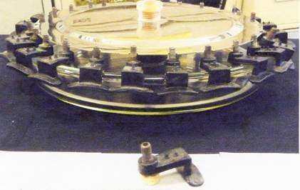
An early EEG machine
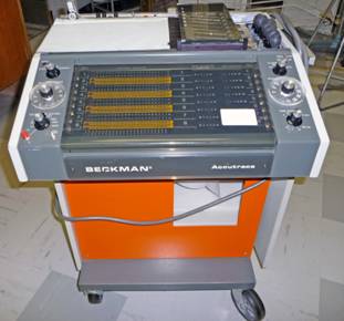
Unlike the current digital models, ink pens were used to produce the recording.
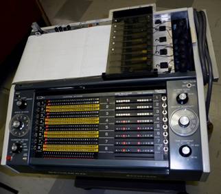 One of the first EEG machines produced by the Beckman companyTop of the machine, showing pens and controls
Early electrodes
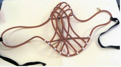
In the current machines the electrodes are an integral part of the skull cap. In the past a string hat was used to hold the electrodes in position.The string hat was placed over the head to hold the electrodes in place. Clip electrodes were placed over the ears and disc electrodes were placed on the mastoid processes behind the ears.
 Chlorinating dish used for the storage of electrodes with a pad electrode in the foreground.
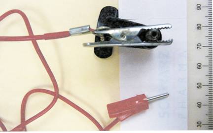 Crocodile ( USA “alligator”) clip attached to the pad electrode. The red cord and the bayonet plug attach it to the EEG machine.
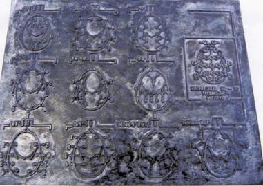 A stencil showing the position of the EEG electrodes.
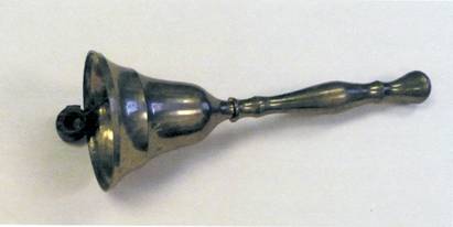 A simple bell was often used to produce the startle reflex as part of the recording validation.
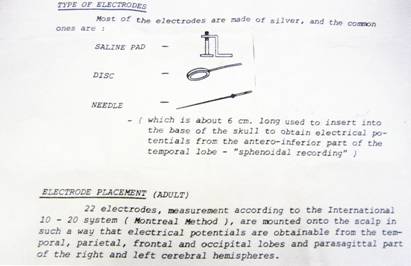 A text book diagram of the commonly used electrodes and a comment on their placements.
Modern electroencephalogram machine
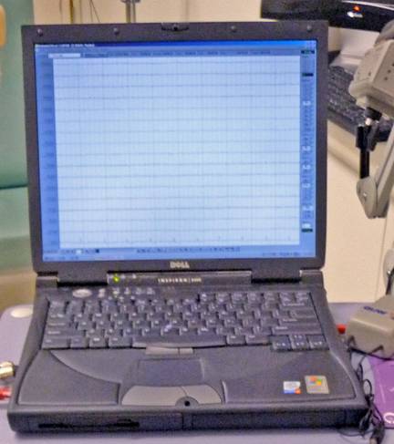
A modern EEG recording station showing 16 simultaneous recording tracings
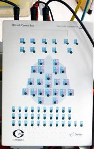
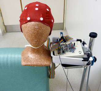 A cap with electrodes as currently used at the QEH. The multicoloured lead connects the electrodes with the relay.... and to the (head) control box below.
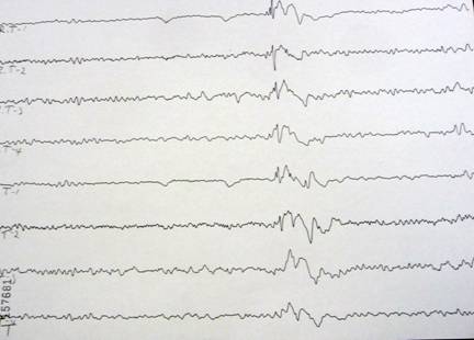 An EEG tracing showing activity in a patient with temporal lobe epilepsy
-o0o-