South Australian Medical Heritage Society Inc
Website for the Virtual Museum
Home
Coming meetings
Past meetings
About the Society
Main Galleries
Medicine
Surgery
Anaesthesia
X-rays
Hospitals,other organisations
Individuals of note
Small Galleries
Ethnic medicine
- Aboriginal
- Chinese
- Mediterran
Dentistry then and nowACKNOWLEDGMENT: The details and photographs have been kindly provided by Professor Wayne Sampson, School of Dentistry, University of Adelaide.
The Oxford dictionary defines a dentist as “one whose profession is to treat the diseases of teeth, extract them, insert artificial ones etc.” Hippocrates and Aristotle (@500 BÇ) mention forceps extractions and the use of wire to stabilise loose teeth or jaw fractures.
The practitioners included priests monks and other healers. These were followed by barbers and the barber’s chair may well have been the precursor of the dental one.
Dental drills originally foot treadle operated (1850) were later provided with electric motors, different drills and faster speeds. Vulcanisation and plastics provided a good material for false teeth. Amalgam fillings, gold tooth crowns and bridges all date back to the 18 th and 19 th centuries, The Adelaide school of dentistry was founded in 1923 and is noted for a major contribution to the development of Orthodontics. Some of the heritage items stored in the museum reflect the development of dentistry in SA
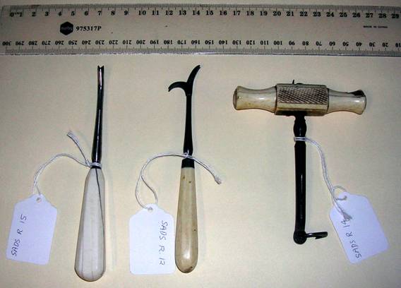
Three late 19th century (1896) dental instruments for the extraction of teeth and roots.
Made from carbon steel with ivory and bone handles.
Early 20th century steel dental forceps
1890 spring form denture (right.) later denture (left.) has a perspex base
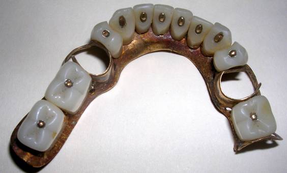
.1860 partial denture constructed from metal and vulcanite held by anchoring studs. The two metal loops were fitted to remaining teeth
Ladle used for heating metal (gold silver etc.) prior to casting
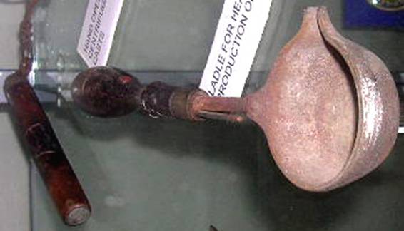
late19 th century centrifugal casting device
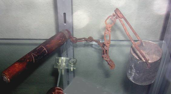
A wax pattern of the restoration to be cast (usually in gold) is inserted in the cylinder with a heat resistant dental plaster (diestone). On heating the wax is lost and molten metal is cast into the die under centrifugal forces created by vigorously swinging the cylinder (crucible) attached to the chain and handle
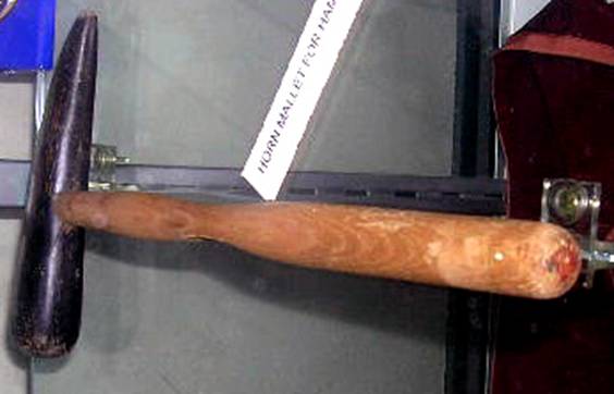
Horn mallet used to hammer soft metal such as gold. (swaging).
Methylated spirit burner used to melt wax or mould plastic
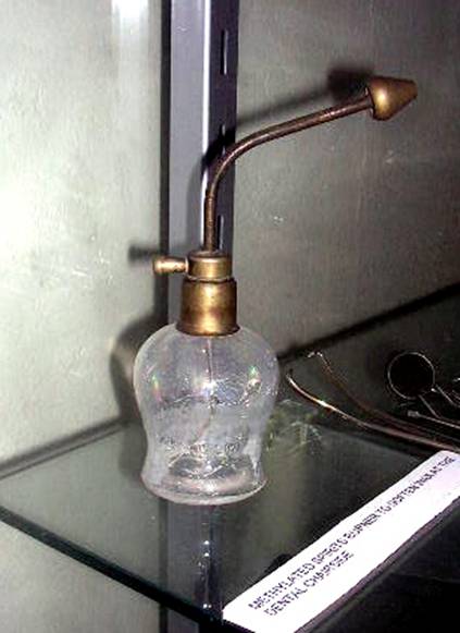
Chip syringe used to puff air to dry surfaces (eg. before placing a tooth restoration
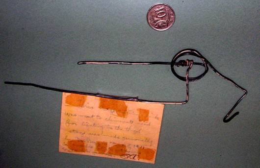
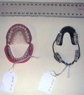
A prototype spring made by Dr. Begg from fencing wire. This was used to show a manufacturer how to design a spring which was used with tooth braces to move tooth roots to correct positions.
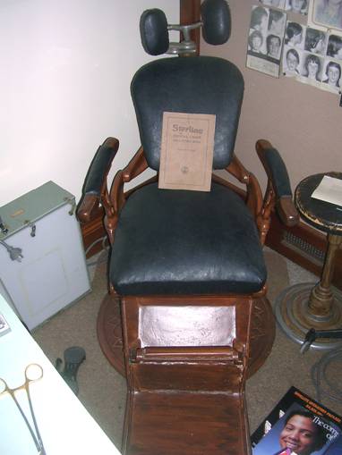
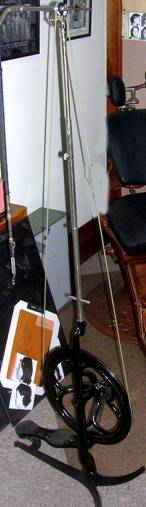
A dental chair & a foot treadle operated dental drill used by Dr. Begg.
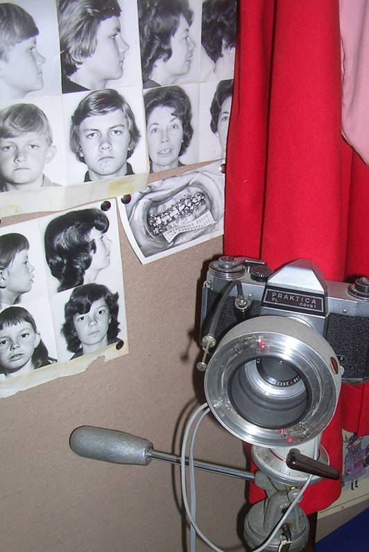
An early “Practica” reflex 35mm camera and circular flash used by Dr. Begg to record photographs before and after orthodontic procedures.
(examples on wall)
Dentistry nowModern technology has provided numerous advances in dental management. Apart from improvements in aesthetic appearance, numerous companies supply equipment which makes dental treatment easier and less painful.
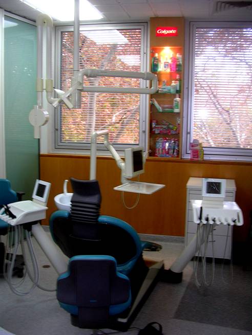
General view of a modern dental suite
Lasers and ultrasound
The use of lasers and ultrasound has revolutionised the treatment of dental caries by improving the cleaning and preparation of dental cavities. Teeth whitening and scaling are performed with ease. Lasers of different wavelengths are used to treat leukoplakia and gum problems. Dental handpieces are air turbine driven and reach speeds of over 200,000 RPM . Their heads can be changed to suit a particular treatment.
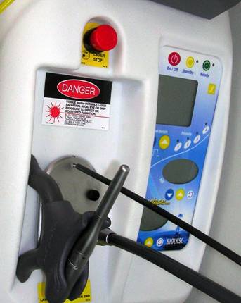
Close-up of laser controls & the probe (center)
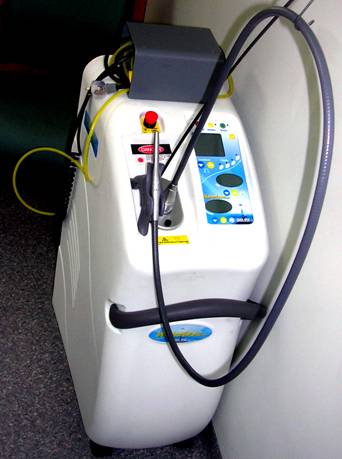
Full view of “Biolase” laser unit.
Computers in dental imaging
Computers are used in dental imaging and in the preparation of dental crowns and bridges, Images of specific teeth are stored on discs and are viewed on a screen attached to the dental chair.
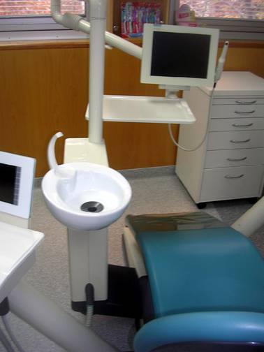
Rinsing bowl and a digital screen for viewing dental x-rays.
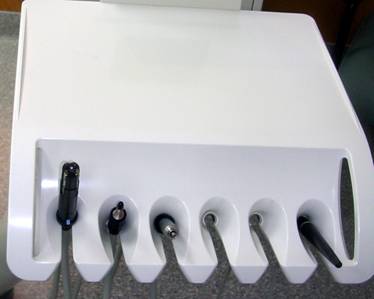
Various dental hand pieces for specific uses
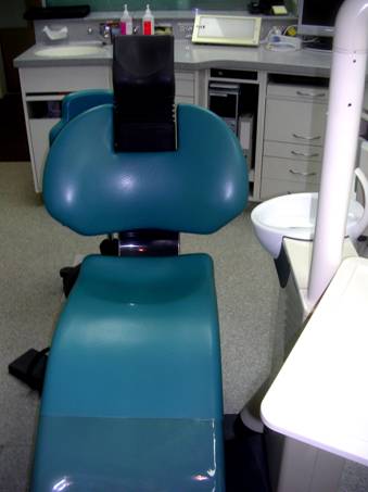
Modern dental chair
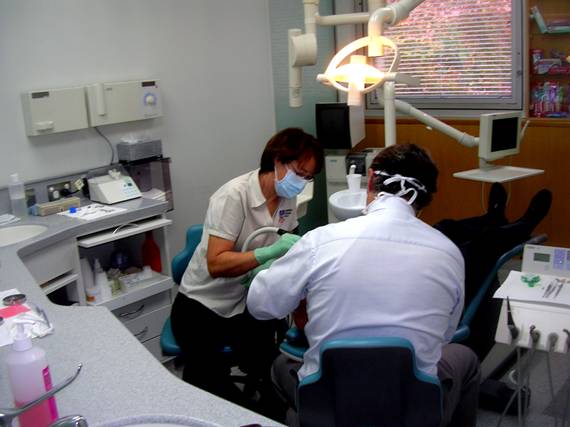
Dentists at work
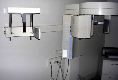
An Orthopantomograph showing head brace and & x-ray unit
The x-ray picture obtained with an orthopantomogram. The larger x-ray film required processing in a radiology department where these machines were usually installed in the past. Modern machines are now installed in the dental suites . Orthodontists can now view an immediate digital image. Note good view of the maxillary sinuses and tempero-mandibular joints.
Caption is below this image
A digital viewing screen which provides immediate pictures of areas of interest. It is no longer necessary to use small x-ray films which had to be placed against the patient’s teeth and then developed, fixed and washed. It has been replaced by a charge coupled device which produces a digital image in the computer. This image is stored and can be retrieved for comparison with a later image.
Caption is below this image
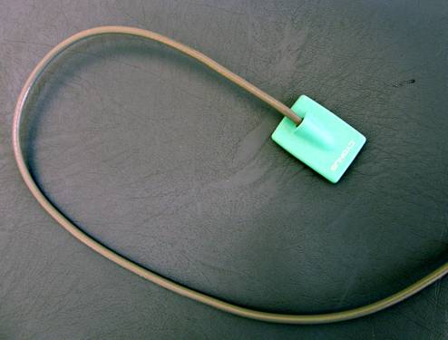
The sensor of a digital dental x-ray unit. It is a very expensive charge coupled device (as in a digital camera) and is placed in the patient’s mouth in place of the now obsolete dental non screen film.
Digital dental x-ray display in a modern dental suite.
Caption is below this image
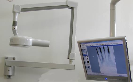
-o0o-A modern x.ray unit is mounted on the wall on the left. The x-ray tube is in the metallic container with a plastic cone which is placed against the patient’s cheek. The digital display is on the right).